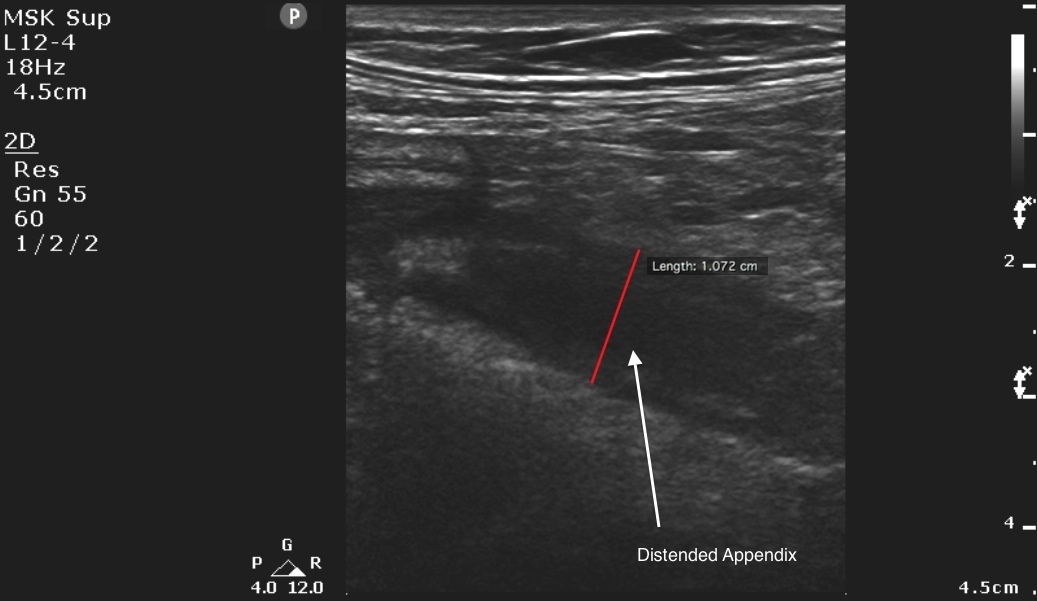
The linear probe is best for evaluating ultrasound in all but the most obese patients. Place the probe at the point of maximal tenderness, which will usually be located around McBurney’s point. The appendix should arise 1 cm below the terminal ileum.
The psoas muscles and the iliac vessels are landmarks anterior to the appendix and may be used to locate the appendix. In normal healthy patients, the appendix is difficult to locate given its smaller size. However, in a patient with appendicitis, the appendix increases in diameter and detection becomes easier.
Criteria for diagnosing appendicitis2:
- Noncompressible, blind-ending tubular structure in the longitudinal axis that measures greater than 6 mm in diameter and lacks peristalsis
- In the transverse view, the distended appendix has a target-like appearance
- Appendicoliths, which appear as hyperechoic foci that cast an anechoic shadow, can also sometimes be found within the lumen of an inflamed appendix
Image 1

Clinical Importance: In addition to being convenient and quick, bedside ultrasound is highly sensitive in the detection of appendicitis.
Literature Support: A meta-analysis by Doria lists the overall sensitivity of ultrasound as 88% and 83% and its specificity as 94% and 93%, for children and adults, respectively.
Doria AS, Moineddin R, Kellenberger CJ, et al. US or CT for diagnosis of appendicitis in children and adults? A meta-analysis. Radiology 2006;241(1):83-94.

