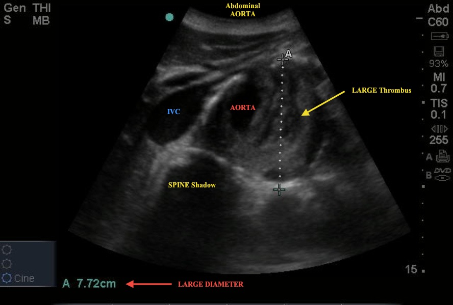
While evaluating a nursing home patient who presented with a hx of CHF and syncopal episodes, the following image was obtained. The wet sounding lungs and CHF exacerbation did not stop the ED evaluation. Bedside US of the abdomen was prompted by a limited history and surgical scars that the patient could not explain.
Note the labeled structures in the image. Also, notice the large mural thrombus and the overall diameter of the aorta - 7.72 cm!
As a reminder, to correctly image the aorta, 5 images should be obtained:
- Proximal Aorta – cross-section with measurement of the diameter
- Mid Aorta – cross-section with measurement of the diameter
- Distal Aorta – cross-section showing bifurcation
- Iliacs – cross-section showing individual diameter measurements
- Longitudinal view – capturing celiac trunk and SMA.


