This patient is a 52-year-old female coming in with chest pain that woke her up and inability to walk.
Bedside ultrasound was done immediately and the images are shown below.
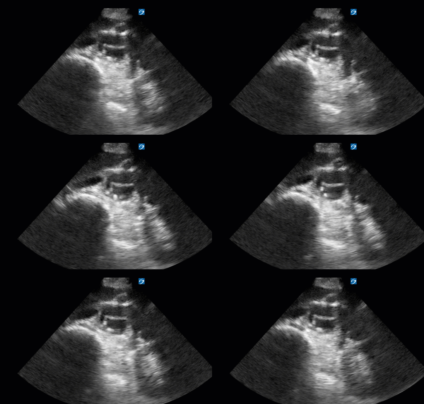
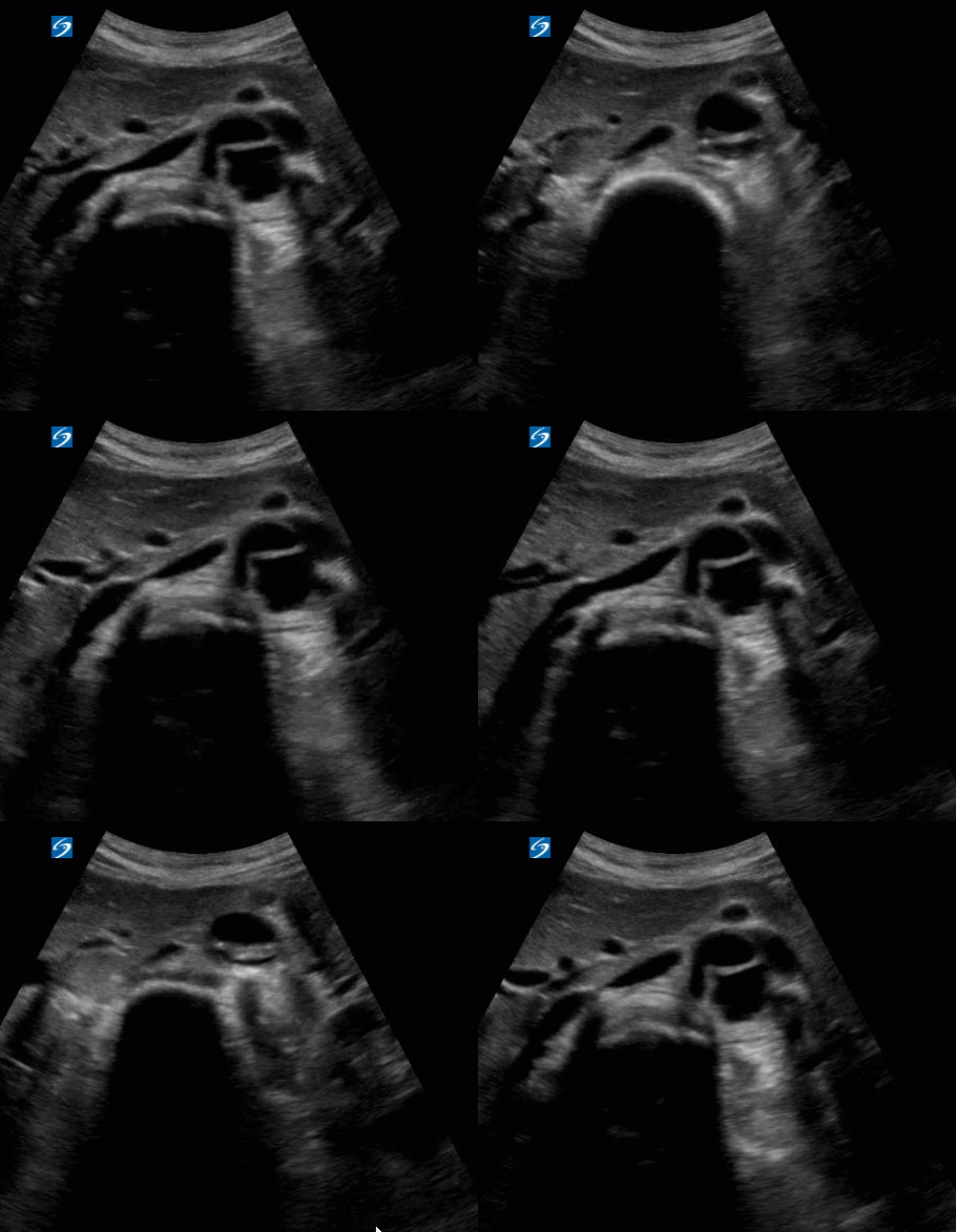
As you can see, there is a view of the dissection flap which CT confirmed as a type A aortic dissection. The patient was on nicardipine and taken to the OR within 70 minutes from arrival to the ED and is currently recuperating in the Grady ICU.
Some tips when scanning the aorta:
Remember to try to find the spine – this is useful in obese patients. With your probe marker to the patient’s right, the aorta should be hugging the vertebral body.
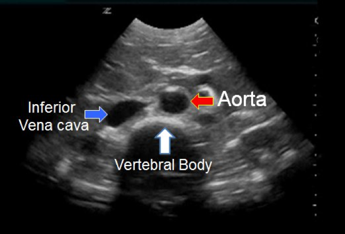
A quick technique is to do a quick pass through and scan from top-down to the iliac bifurcation to look for any abnormalities.
Keep the vertebral body in view and remember to save images of the proximal aorta, middle aorta, and distal aorta. You may not always see a flap but you can pick up an aneurysm. Measure the outer wall to outer wall.
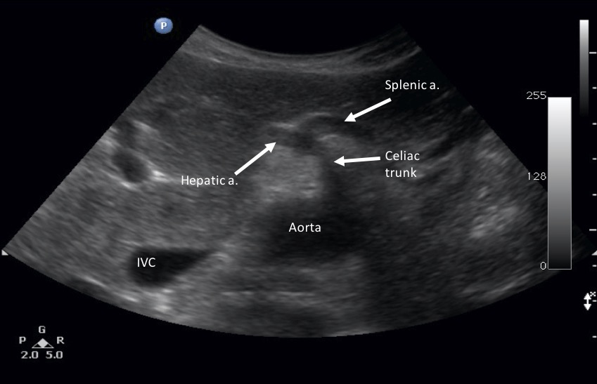
Proximal Aorta - you can see the seagull sign (splenic and hepatic artery bifurcation off the celiac trunk). This view may be more challenging in larger adults.
Middle Aorta
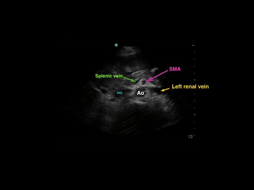
Distal Aorta
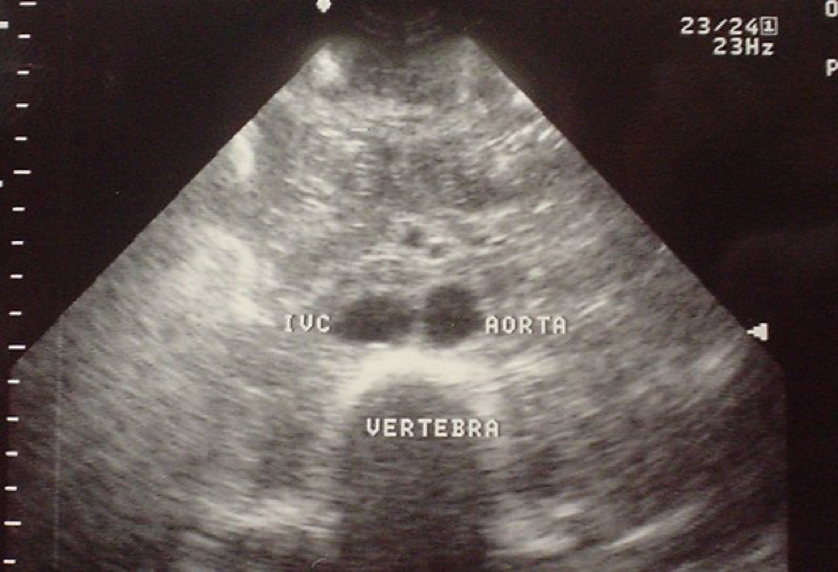
Keep in mind that the overall sensitivity of bedside ECHO is only 59-83%. It is difficult to pick up an intimal flap and extremely difficult to get a view of the aortic arch. (See here for the suprasternal notch view).
You can arrange for an emergent TEE if the concern is high as the sensitivity of a TEE in diagnosing dissection is 85-90%. However, keep in mind there is also a 2-cm blind spot proximal to the innominate arteries.
CT sensitivity is around 94% and MRI is 95%.
Date: 2018

