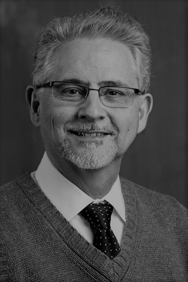
Ernest Garcia joined Emory Radiology in 1985 as an associate professor of radiology in 1985, received tenure in 1987, and was promoted to full professor of radiology in 1990. His innovations in the field of nuclear cardiology fueled his rapid ascent.
Dr. Garcia’s early realization that interpretation of nuclear medicine images lacked reproducibility and objectivity drove him to apply his computer programming expertise as well as his training in nuclear physics to transform the art of image interpretation into the science of image interpretation. His landmark article, ‘‘Quantification of rotational thallium-201 myocardial tomography,” did just that. It built on his earlier research using interpolative background subtraction, circumferential count profiles, display of spatial distribution of Thallium-201, and changes of images over time through redistribution to quantitatively analyze planar myocardial perfusion images.
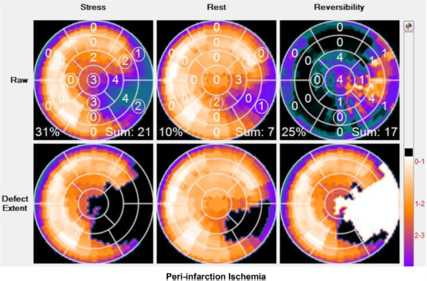
The excited response to his 1985 publication inspired Dr. Garcia to expand the software training database with data from thousands of patients with heart disease as well as normal subjects. The software then was able to discern what was within normal range and qualify what was clinically significant. The result: the Emory Cardiac Toolbox TM, one of the earliest artificial intelligence (AI) clinical decision-making tools providing a standard approach for image processing.
Emory University licensed the first of Dr. Garcia’s software prototypes in 1993 and licensed the first version of the complete Emory Cardiac ToolboxTM in 1999 to General Electric, Siemens, Philips and Syntermed. Over the years, numerous clinical trials validated the computer software as producing accurate diagnoses faster than live physicians. For every new advance in cardiac imaging, Dr. Garcia and his team developed corresponding new tools for the growing Emory Cardiac ToolboxTM. Other inventors followed Garcia’s lead, but the Emory Cardiac ToolboxTM continues to be used worldwide for processing millions of imaging tests annually.
Dr. Garcia introduced numerous innovations in the field of cardiac imaging. They include quantification of planar, SPECT and PET myocardial perfusion imaging; fusion, quantification and visualization of multimodality cardiac imaging, specifically perfusion and coronary anatomy; quantification of left ventricular dyssynchrony; expert systems for aiding cardiac and renal imaging interpretation; and the development of the real-time 2D echo computer interface.

In 2008, Dr. Garcia suffered a cardiac event himself. Like millions of other patients, his doctors turned to the Emory Cardiac ToolboxTM to interpret his images. His own invention showed the presence of coronary artery disease and myocardial infarction. This became part of a story in Newsweek magazine about researchers who get diagnosed with their own inventions.
During his career Dr. Garcia received many awards and honors, including the Ernest V. Garcia, PhD Endowed Professorship in Cardiac Imaging in recognition of his impact on the field of cardiac imaging to the benefit of countless patients worldwide.
He has served in many national leadership positions. He served as president of the Cardiovascular Council of the Society of Nuclear Medicine (1990), the Institute for Clinical PET (1996), the American Society for Nuclear Cardiology (2002), as founding president of the American Society for Nuclear Cardiology Foundation (2003). He served on the Editorial Boards of most premier medical and cardiac imaging journals and was named one of the Top 10 Nuclear Medicine Researchers by the readers of Medical Imaging Magazine (2005 and 2006). He also received honors from the Better World Project for Academic Research that Changed the World (2008).
Emory honors include the Emory University Top Innovator Award (2007) and the Emory University School of Medicine Game-Changer award for his role in the development of tools for the objectification of image interpretation (2012).
Ernest Garcia is author or co-author of more than 400 original articles, editor or co-editor of 10 books, and author of 65 book chapters. Upon retirement in 2023, Dr. Garcia said his greatest satisfaction is walking into a hospital anywhere in world and seeing his software up and running and affecting patient outcomes.
Dr. Garcia was recruited by the University of Miami while he was a senior in high school and working as a salaried computer programmer for a local company and offered a full college scholarship to study physics. After earning his undergraduate degree, he received a full fellowship in physics with additional stipends to pursue a PhD in astrophysics. During the first year of his doctoral program in astrophysics, NASA’s annual budgets were drastically cut, and a friend suggested he explore a relatively new field called nuclear medicine. He decided to apply his training in physics and software development to improve clinical medicine and earned his PhD with a dissertation on attenuation correction of scintillation camera images.
He began his academic career as an instructor in radiology at the University of Miami School of Medicine in 1974. After quickly rising through the ranks to the level of associate professor, he joined the nuclear cardiology group at Cedars-Sinai Medical Center as a collaborator in 1979 and became the director of computer sciences in 1980. In 1983 he was appointed adjunct associate professor of radiological sciences, Cedars-Sinai Medical Center, University of California at Los Angeles (UCLA).
Upon retirement in 2022, Dr. Garcia said his greatest satisfaction is walking into a hospital anywhere in world and seeing his software up and running and affecting patient outcomes.

