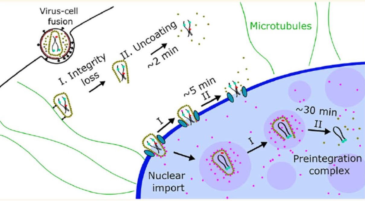
Timely disassembly of the cone-shaped HIV-1 capsid/core (uncoating) after virus-cell fusion is a prerequisite for establishing infection. We have visualized HIV-1 uncoating by indirectly labeling the viral capsid with CypA-DsRed and directly labeling through amber codon suppression. Co-labeling click-labeled CA protein with organic dyes and trapping a fluid-phase marker within the core enables monitoring the uncoating intermediates, from small defect formation to core disassembly (Figure 2 and Movie 2). This co-labeling approach revealed intact HIV-1 cores entering and uncoating in the nucleus. We also found that viral pre-integration complexes are transported to nuclear speckles in macrophages and other cell types where they integrate into speckle-associated genomic domains, SPADs.


